Case Study: ACL Reconstruction using Quad Tendon
Autograft andMeniscus Repair in a 25-year-old Female
A 25-year-old female injured her left knee during a soccer game a month ago. She felt a snapping sensation in her left knee while playing soccer. The injury was followed by knee pain and swelling. The swelling subsided over the last month but the patient reports a sensation of her knee giving away during her day-to-day activities.
The patient works as an UPS driver but is currently not working secondary to the knee injury. The patient reports pain and a feeling of her knee giving away while walking, climbing stairs, and turning. The patient’s medical history is significant for polycystic ovarian syndrome and is currently on oral contraceptive medications. She is a current smoker and denies any illicit drug use.
On physical examination of the left knee, the effusion has significantly subsided compared to her visit at the time of the injury. Anterior drawer test and Lachman test are positive (2+). Mcmurray’s test for medial and lateral meniscus is positive. The range of motion of the left knee is limited secondary to pain (flexion 80 degrees and extension 15 degrees).

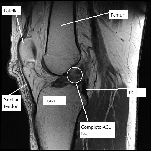
MRI of the left knee showing complete tear of the ACL.
Radiological study in the form of an MRI suggested a complete tear of the ACL along with complex tears of both the medial and the lateral meniscus. The patient desired to be athletically active and to continue her job without any episodes of instability.
Various treatment options were discussed for the patient’s diagnosis, which included: living with the extremity as it is, organized exercises, medicines, injections, and surgical options. The patient has expressed a desire to proceed with surgery. The risks, benefits, and potential complications were all discussed with the patient at length.
The patient also quit smoking before the surgery with a plan not to start it after the surgery, considering our discussion with regards to detrimental effects of smoking on healing and high risk of failure..
PREOPERATIVE DIAGNOSES:
- ACL tear of the left knee.
- Medial meniscus tear of the left knee.
- Lateral meniscus tear of the left knee.
POSTOPERATIVE DIAGNOSES:
- ACL tear of the left knee.
- Medial meniscus tear of the left knee.
- Lateral meniscus tear of the left knee.
OPERATION:
- Left knee arthroscopic medial meniscus repair.
- Left knee arthroscopic lateral meniscus repair.
- Left knee arthroscopic ACL reconstruction using quad tendon autograft.
PROCEDURE: The patient was taken to the operating room where he was placed on a well-padded operating table. General anesthesia was induced. Adductor canal block was given. A mid-thigh tourniquet was applied. Left lower extremity was prepped and draped in the usual fashion.
A timeout was called. The preop antibiotic was given. Esmarch was used to exsanguinate the limb and the tourniquet was elevated to 300 mmHg. The timeout was called.

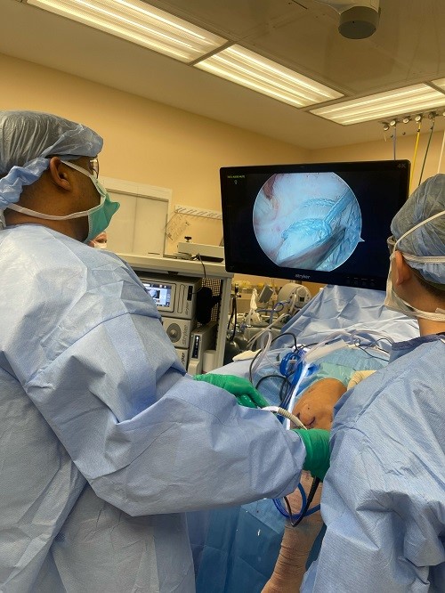
Intraoperative arthroscopic image of the left knee.
A lateral entry portal was made and an arthroscope was introduced into the knee joint. There was no osteochondral lesion of the patellofemoral joint, medial condyle, lateral condyle. Examination of the medial meniscus showed a mid body oblique vertical tear of the medial meniscus along with a tear of the posterior horn of the medial meniscus.
There was an ACL tear, which was not repairable due to it being midsubstance. There was a lateral meniscus tear along with the mid substance and anterior body of the lateral meniscus, which was flipped inside.
A Medial entry portal was made. The probe was introduced and the lateral meniscus tear was reduced with difficulty. The lateral meniscus tear was held in place using a spinal needle, through which a FiberWire was passed. The FiberWire was used as a holding suture. Meniscal stitcher from Arthrex were used x 2 to repair the lateral meniscus. Similarly, the medial meniscus was also reduced and sutured using two meniscal stitches from Arthrex. The repairs were satisfactory. Pictures were taken and saved..

Intraoperative picture showing knee arthroscopic surgery.
Now, the quadriceps tendon graft was harvested through a 3-cm anterior midline prepatellar incision. The fascia was cut in the line of the incision. The quadriceps tendon was identified. A 10 mm double blade with 10 mm depth was used to cut the tendon in between vastus medialis and vastus lateralis to a depth of 80 mm.
The undersurface of the tendon was freed by sharp dissection. Closed quadriceps tendon cutter was used to harvest the tendon. The tendon was prepped on the back table and FiberWire with the FiberTag was used to prep each end.
The length of the graft was 80 mm. The graft was marked at 35 mm on the femoral side. The intercondylar notch was prepped for ACL insertion with the use of shaver. The entry portal was made using the ACL guide.
The lateral exit point was marked through the guide and incision given. The ACL femoral guide was inserted and a FlipCutter was inserted from the lateral condyle towards the femur inside a drill sleeve. When it came into the medial end of lateral condyle and seen arthroscopically, the cutter was flipped.
About 30 mm tunnel was made on the femoral side with the FlipCutter. Similarly, the tibial footprint was cleaned and a tibial tunnel was made with an ACL tibial guide using the flip cutter.
A passport cannula was inserted on the medial portal for easy suture management and passage of the graft. Once the tunnels were done, FiberLoop was passed on either side. The femoral side was brought out of the passport with TightRope was passed and passed through the femoral tunnel under arthroscopic visualization.
Once 25 mm was crossed, the tibial loop was extracted and the tibial TightRope was passed. Once the tibial TightRope was out of the bone, the button was flipped and tightened. Similarly, the button was flipped on the femoral side and tightened. The sequential tightening was done on the tibial and femoral sites for the TightRope.
The knee was cycled 30 times into flexion and extension. The final tightening was again performed in extension on the femoral and tibial side and the TightRopes were tied over each other.
The wounds were thoroughly washed and irrigated, all the debris was removed with the shaver. After proper irrigation, the knee was irrigated and closed using #3-0 nylon. The quadriceps tendon incision was irrigated and closed in layers using #2-0 Vicryl and #3-0 Monocryl.
The wounds were dressed using 4 x 8s, ABDs, Webril, and Ace wrap. The Bledsoe knee immobilizer was advised. The patient was extubated and moved to recovery in stable condition. The patient’s neurological status was intact after the surgery.
Disclaimer – Patient’s name, age, sex, dates, events have been changed or modified to protect patient privacy.
My name is Dr. Suhirad Khokhar, and am an orthopaedic surgeon. I completed my MBBS (Bachelor of Medicine & Bachelor of Surgery) at Govt. Medical College, Patiala, India.
I specialize in musculoskeletal disorders and their management, and have personally approved of and written this content.
My profile page has all of my educational information, work experience, and all the pages on this site that I've contributed to.

