Case Study: Shoulder Scope: Labral Debridement
Rotator Debridement, ACR, DCR in a 36 year-old male
Many labral rips are treated with an arthroscope. If the tear is tiny and largely getting caught as you move the shoulder, merely removing the ragged edges and any loose labrum fragments may relieve your problems.
Debridement is the removal of loose tendon pieces, thickened bursa, and other material from around the shoulder joint. To alleviate chronic shoulder pain, shoulder surgeons undertake a Distal Clavicle Resection operation.
For the past few months, a 36 year-old male patient came to the office due to right shoulder pain. We performed an MRI, which revealed disease in the subacromial and labrum regions. The AC joint is also sensitive for the sufferer. The patient received conservative treatment, but no relief was provided.
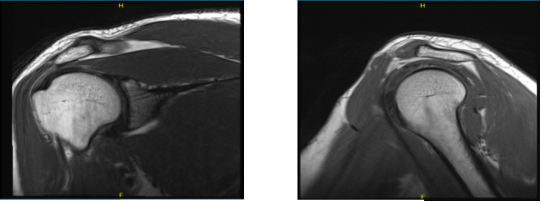
MRI findings; Evaluation of the regional osseous structures shows no evidence of marrow edema or contour deformity to suggest bony injury. There is nonspecific subchondral cyst formation at the poster superolateral humeral head. The glenohumeral and acromioclavicular joints are normally aligned.
The acromial process is posterolateral down sloping and its undersurface is generally straight in configuration. The coracoacromial arch closely approaches the underlying supraspinatus muscle/tendon, which may manifest clinically as subacromial impingement.
There is a tear of the inferior insertional fibers of the infraspinatus tendon. The supraspinatus, subscapularis, and teres minor tendons are unremarkable in appearance. There is a partial tear/tendinopathy of the intra-articular biceps’ tendon.
The intertubercular segment of the biceps’ tendon demonstrates normal course and morphology. There is superior labral fraying, as well as stripping of the superior labrum-biceps tendon anchor from the supraglenoid tubercle. This is compatible with a SLAP (superior labrum anterior-posterior) lesion.
The remainder of the glenoid labrum is intact. The regional musculature is well-developed, without evidence of edema or atrophy. There is a small amount of physiologic fluid within the glenohumeral joint.
There is no fluid within the subacromial-subdeltoid or subcoracoid bursae. Incidental note is made of a subcutaneous lipoma within the soft tissues at the posterosuperior aspect of the acromion process. It measures approximately 2.5 cm in greatest dimension.
Despite our discussions of different therapy choices, the patient chose surgical intervention. We talked about the need for physical treatment and rehabilitation.
We talked about, among other things, the necessity for rehabilitation, harm to nearby nerves and muscles, tingling, numbness, and ecchymosis. The client was aware and gave informed consent.

MRI-3T Right shoulder non-contrast
The patient was brought into the operating room and set down on a well-padded operating table. Anesthesia was induced throughout. The right shoulder was raised as the patient was positioned in a left lateral position. The right shoulder was prepared as normal and wrapped aseptically.
On a 10-pound weight traction, the right shoulder was positioned in 10 degrees of flexion and 45 degrees of abduction. A timeout was ordered. An antibiotic was given before surgery.
Through a soft area, the posterior entry hole was created. Through the posterior portal, the arthroscope was inserted into the glenohumeral joint. When the glenohumeral joint was examined, it was discovered that both the superior and posterior labrums were torn.
Additionally, it revealed a partial rotator cuff from the supraspinatus on the articular side in the area of the rotator interval. Debridement was performed to remove the rotator cuff and the superior labrum’s fraying and tear. The rest of the shoulder was examined, and everything was fine. Images were captured and stored.
The anterior portal was used to introduce the arthroscope, which was then used to confirm the results and perform debridement. The anterior surface of the acromion and the CA ligament were seen to be fraying as the arthroscope was inserted into the subacromial region.
A thermal wand was used during the acromioplasty procedure, followed by a bur. The ligament of the CA was freed. When the AC joint was revealed, degenerative changes were discovered. Utilizing the heat wand and the bur through the anterior and posterior portals, the distal clavicle was excised. The clavicle’s top centimeter was removed. Final images were captured and stored.
The shoulder was irrigated and drained carefully. The arthroscopic incisions were made with #4-0 nylon. Dressing was completed with. The patient’s arm was slung over his shoulder. In a stable state, the patient was extubated and transported to the recovery unit.
The patient seen in the office for his post-operative consultation, he denies fever and chills. He has occasional pain in right shoulder though he is able to move the shoulder.
He has almost full ROM and strength and is working with his PT and its improving cause now he is able to perform all usual activities and can return back to work. Upon examination, we agreed to proceed with formal physical therapy as well as a home exercise program, use ice, elevation and NSAID’s for pain PRN for shoulder.
The patient did well after the surgery and with the help of physical therapy, he is improving, gradually. The doctor advised the patient to visit the office whenever he needed to.
Disclaimer – Patient’s name, age, sex, dates, events have been changed or modified to protect patient privacy.
I am Vedant Vaksha, Fellowship trained Spine, Sports and Arthroscopic Surgeon at Complete Orthopedics. I take care of patients with ailments of the neck, back, shoulder, knee, elbow and ankle. I personally approve this content and have written most of it myself.
Please take a look at my profile page and don't hesitate to come in and talk.

