Case Study: Microdiscectomy (Right) L4-L5 in a 58-year-old Female
The patient is in our office, referred by her primary care physician with complaints of right lower back and right leg pain along the front of the knee, leg, and dorsum of the foot for the last 3 months. The patient remembers of an injury when she was moving stuff from the attic at home.
The pain is extremely severe in intensity (9/10 on a scale of 0 to10). The patient describes the pain as sharp, stabbing, burning. The pain is constant and does not disturb sleep. The pain is associated with tingling, numbness, radiating pain, limping. The patient’s torso is bent to left due to pain and he walks with a discomfort.
The pain is not associated with swelling, bruising, bowel or bladder abnormality, hand function difficulty. The patient’s symptoms had worsened over time. Activities such as walking, standing, lifting, exercise, twisting, lying in bed, bending, squatting, kneeling, stairs, sitting make the symptoms worse. Rest, heat, ice, lying down, bending forwards makes the symptoms better.
He has tried medications and rest along with therapy treatment as per his PCP but has had no relief. The patient is currently working as an assistant in a dental office. The constant pain has affected her daily activities and she is currently out of work secondary to pain. The patient does not smoke, is on no medications, and has no allergies.
On general physical examination, the patient is calm, conscious, cooperative, and well oriented to time place and person. On examination of the lumbar spine, the patient is tender to palpation over the paraspinal musculature. There is no tenderness over the spinous processes and there is no crepitus with ranging.
The range of motion of the lumbar spine was limited due to discomfort. An obvious sciatic list to the left side is present. There is a positive straight leg raise test on the right at 30 degrees and contralateral of the left at 40 degrees. There is no tenderness to palpation over the trochanteric bursa and hip.
There is no soft tissue swelling and ecchymosis. The patient has a full range of motion of the hips. The hips are stable on the examination. There are no erythema, warmth, or skin lesions present.
Range of motion:
Flexion (Normal – 60 degrees) – 40
Extension (Normal – 45 degrees) – 30
Lateral Flexion Right (Normal – 45 degrees) – 30
Lateral Flexion Left (Normal – 45 degrees) – 30
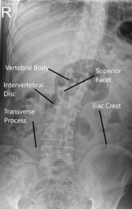
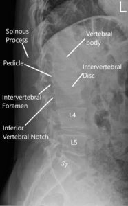
X-ray of the LS spine suggested degenerative changes and moderate dextroscoliosis.

LS Spine X-ray in AP and Lateral views.
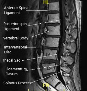
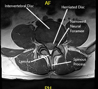
MRI of the LS spine suggested a posterior extruded disc herniation at the L4-L5 level causing severe spinal canal stenosis and compression of descending L5 nerve roots. Right paramedian posterior disc herniation L4-L5 level causing mild spinal canal stenosis and degenerative changes.

MRI of LS spine in sagittal and axial sections.
Her pain and weakness had been worsening, so she presented to the hospital where she was admitted for the same. Her Imaging studies were reviewed and discussed with the patient at length.
We discussed treatment options and the patient opted for surgical management. Risks and complications Including Infection, bleedIng, Injury to nerves and vessels, aggravation of symptoms, aggravation of pain, future instability, and need for future surgery, back pain, rehabilitation among others. The patient understood and signed the informed consent.
PREOPERATIVE DIAGNOSES: Extruded disc L4-5 right side with radiculopathy and weakness of right lower extremity.
POSTOPERATIVE DIAGNOSES: Extruded disc L4-5 right side with radiculopathy and weakness of right lower extremity.
OPERATION: Microdiscectomy, right L4-5.
DESCRIPTION OF PROCEDURE: An incision was given in the midline over L4-5. Right-sided dissection was performed on the inferior lamina of L4 and superior lamina of L5 and the spinous process. The levels were again confirmed under C-arm and the picture was saved.
The microscope was brought in. The laminotomy of L4 was performed under a microscope using an electric matchstick burr. The ligamentum flavum was raised. The flavum was excised and foraminotomy was performed. The dural sac and the nerve root were retracted medially.
Bipolar cautery was used to achieve hemostasis. The disc space was reached at the L4-5 level and found to be having a deficiency in the posterior longitudinal ligament.
The extruded fragment was found just anterior to the thecal sac. The fragment was removed. Discectomy was performed and washed thoroughly. Satisfactory decompression as well as foraminotomy was achieved. Visipaque dye was used to confirm the level as well as decompression.
Final pictures were again saved. The dye was washed off. Hemostasis was finally achieved and Surgiflo mixed with 40 mg of Depo-Medrol was instilled. One gram of vancomycin was also instilled in the wound.
The wound was closed In layers. The patient was turned supine In the bed and extubated and moved to the recovery unit In stable condition.
POSTOPERATIVE EXAMINATION: The patient was neurologically intact. His pain was decreased in his right lower extremity. He was complaining of mild pain in his back
Disclaimer – Patient’s name, age, sex, dates, events have been changed or modified to protect patient privacy.
My name is Dr. Suhirad Khokhar, and am an orthopaedic surgeon. I completed my MBBS (Bachelor of Medicine & Bachelor of Surgery) at Govt. Medical College, Patiala, India.
I specialize in musculoskeletal disorders and their management, and have personally approved of and written this content.
My profile page has all of my educational information, work experience, and all the pages on this site that I've contributed to.


